Advanced Image Recognition Technology
Cutting Edge Science; Breakthrough Technology
ProMark uses highly-intelligent and automated imaging technology. This advanced technology allows ProMark to deliver focused and precise image analysis, and develop a rapid, completely objective, and fully reproducible result to inform your decision-making process.
ProMark’s technology platform measures the exact amounts of multiple protein biomarkers on standard formalin-fixed paraffin-embedded tissue sections, in highly defined sub-regions (“regions of interest”) of intact tissue where ProMark-specific biomarkers are known to be altered as part of prostate cancer processes. Learn more.
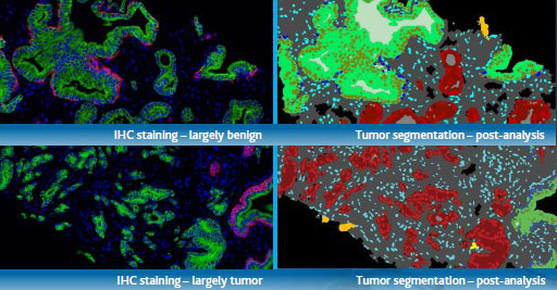
Protein biomarkers are measured in situ
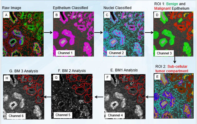
- The raw image (A) contains spectral information from the tumor mask (cytokeratin), DAPI, and prognosis determinants.
- The algorithm classifies stroma, background, and epithelium based on markers for cytokeratin (B), and classifies nuclei based on DAPI (C).
- Nuclei are further classified into stromal, malignant, or benign based on additional tumor mask (D) and then glands are classified as either benign or malignant based on the nuclei and morphologic features (E).
- This ultimately results in several specific regions of interest for biomarker measurement (F).